CryoFlash · Time-resolved cryo‑EM

Microsecond time-resolved cryo‑EM by melt and freeze

CryoFlash is a benchtop instrument that melts and re-freezes a vitrified cryo‑EM sample in microseconds, in regions that you select. While the sample is liquid, proteins are made to perform their tasks. Re-freezing then traps their motions for standard cryo‑EM imaging.

External testing at the DCI Lausanne in 2026. First instruments will be available starting 2027.

What is CryoFlash?

Trigger protein dynamics. Capture the moment.

CryoFlash uses precisely controlled laser pulses to melt and re-freeze regions that you select on a vitrified cryo‑EM sample. While the sample is liquid, a stimulus of your choice induces protein dynamics. When the laser stops, the sample re-freezes and the protein motion is trapped. Different regions can capture different time points on the same sample.

Explore the science · Read the publications

  • Works with cryo‑EM samples on metal support films, for example UltrAuFoil
  • Calibrated, reproducible melt–freeze cycles
  • Returns a sample ready for standard cryo‑EM imaging

Capturing a virus in motion

Published research using the melt-and-freeze method behind CryoFlash captured a virus capsid partway through a structural transition.

A virus that has to change shape

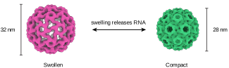
Cowpea chlorotic mottle virus (CCMV) packages its RNA genome inside a capsid of 180 proteins. In its compact, 28-nanometre form the capsid protects the genome; to release the RNA, it swells to 32 nanometres. The endpoints were known; the challenge was to capture the fleeting structures that connect them.

The CCMV capsid in its compact 28 nanometre form and its swollen 32 nanometre form, side by side.

Two shapes, and a gap

Conventional cryo‑EM samples reveal the compact and swollen populations. The missing information is what happens during the short transition between them.

A plot of particle populations showing two clusters: swollen capsids at 32 nanometres and compact capsids at 28 nanometres, with empty space between them. A third cluster between the other two: capsids caught mid-collapse at 31 nanometres.

Conventional cryo‑EM

Intermediate captured by melt and freeze

3D variability analysis of the particle populations. Components 1 and 2 are the two directions in which the particles differ most from one another. Every particle takes a position along them, so particles of similar shape land in the same spot, and the shading shows how many of them sit there. The blue population contains particles caught partway through the transition.

Caught in the middle

The laser kept the sample liquid for thirty microseconds (0.000030 seconds), long enough for the capsids to begin collapsing before they were frozen again. The result was a third population: particles caught partway, no longer swollen and not yet compact. CryoFlash brings this melt-and-freeze approach into a dedicated benchtop instrument.

The pH stimulus was prepared while the sample was frozen; the virus could respond only once the film became liquid. More about trigger timing.

Atomic models fitted to reconstructions along the contraction pathway, interpolated between them.

Figures adapted from Harder et al., Nature Communications 14, 5649 (2023), published under CC BY 4.0. Axes and labels simplified; maps low-pass filtered to a common resolution for display.

How CryoFlash fits into the workflow

One new step in a workflow you already know

CryoFlash sits between vitrifying and imaging. Insert your vitrified sample into CryoFlash before going to the transmission electron microscope for imaging. A key requirement is a metal sample-support film, for example gold, silver, or copper, so the melted spot can re-vitrify.

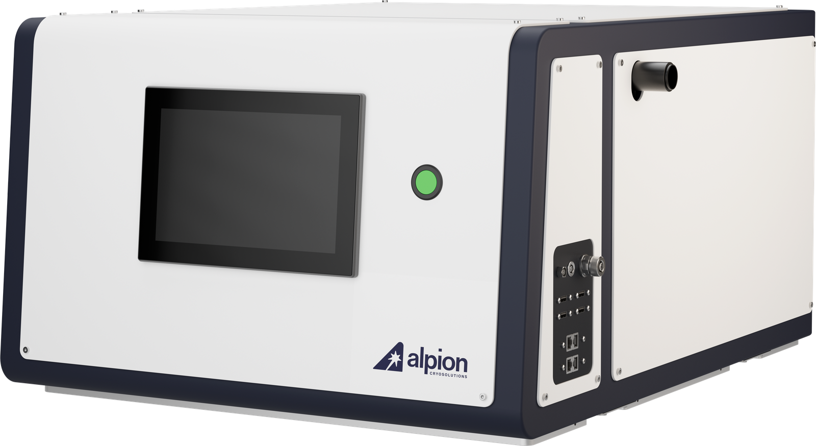
The CryoFlash benchtop instrument, a table-top melt and freeze system
The CryoFlash instrument: melt and freeze, on the bench. Product visualization.

Start with your purified sample

Start with the sample you want to study.

Vitrify your sample as usual

Use your usual plunge-freezing setup with a metal sample-support film that conducts heat away rapidly enough for revitrification.

CryoFlash Our instrument

Melt, move, re-freeze in microseconds

Calibrated melt-and-freeze cycles capture transient protein structures across a series of delays: the frames of a molecular movie.

Image on your own microscope

Collect standard cryo‑EM images on your existing microscope, targeting the revitrified regions.

Process & reconstruct

Reconstruct the captured states and analyze the structural changes along the pathway of the motion.

The workflow

From grid loading to a recorded melt-and-freeze experiment

Load a grid, map it, select target areas, and run calibrated melt-and-freeze cycles. Then transfer the grid to your microscope for data collection.

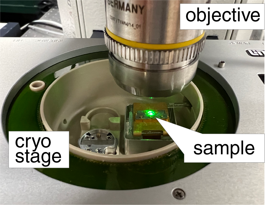
1 A vitrified sample loaded on the cooled CryoFlash cryo-stage beneath the optical objective

Load the grid

Place your vitrified sample into the cooled cryo-stage, ready for optical imaging.

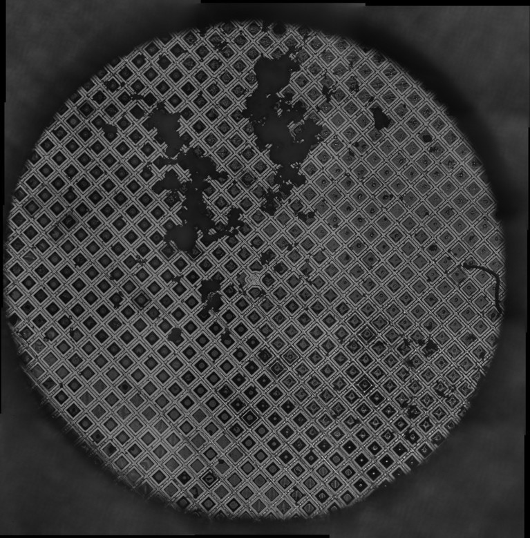
2 Whole-grid atlas montage acquired by the optical microscope

Map the grid

Acquire an optical atlas of the whole grid to find the regions best suited to melt and freeze.

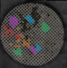
3 Atlas with several target grid squares highlighted for melt and freeze

Choose grid squares

Select the squares to act on, and the software runs the experiment on every target precisely.

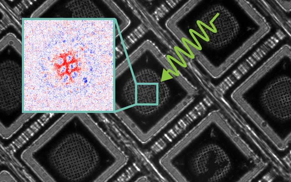
4 Laser melt and freeze of a single grid square, with diffraction inset

Melt & freeze

A calibrated laser pulse melts and re-vitrifies each square, trapping transient states, and the software logs every one into an interactive atlas.

5

Transfer to TEM

Transfer the grid to your electron microscope and image it like any other cryo‑EM sample, using the interactive atlas to set up data collection.

CryoFlash software guides grid mapping, target selection, and calibrated melt-and-freeze experiments. Once configured, the melt-and-freeze series runs unattended. An interactive atlas records the settings and outcome for each treated square and helps guide subsequent data collection. Have a question about your application? Talk to us.

System overview

CryoFlash at a glance

The essentials of CryoFlash and its workflow. Contact us to discuss the configuration and requirements for your laboratory.

Method
Laser melt and freeze of vitrified cryo‑EM samples
Time resolution
A few microseconds, depending on the support-film type
Input
Standard plunge-frozen cryo‑EM sample on a metal support film, for example UltrAuFoil
Output
A sample ready for standard cryo‑EM imaging
Targeting
Software-guided square selection on an optical atlas
Automation
Software-guided mapping and unattended melt-and-freeze series
Format
Benchtop
Footprint, power, and laser class
On request
Status
External testing at the DCI Lausanne in 2026. First instruments will be available starting 2027.

Get started

Join the CryoFlash waiting list

The waiting list is non-binding: no payment and no purchase commitment. Tell us about the samples you work with and we will follow up to discuss technical fit, the current validation status and expected availability.

The science

Time-resolved cryo‑EM by melt and freeze

Conventional cryo‑EM reveals structures preserved at vitrification. Melt and freeze adds control over the brief interval in which proteins can move before their structures are trapped again.

A cryo‑EM sample is first vitrified by the standard route. A brief, precisely controlled laser pulse heats the metal support film, which in turn melts the vitreous film for a few tens of microseconds, long enough for the proteins inside to move. The surrounding support, held at cryogenic temperature, then pulls the heat away and re-vitrifies the film almost instantly, trapping the transient state that had been reached. Collecting particle images across a series of delays turns the reconstructions into frames of a molecular movie with microsecond time resolution. In the scientific literature the approach is known as melting and revitrification; on this site we simply call it melt and freeze.

The melt-and-freeze cycle in four steps: a laser pulse melts the vitreous film from 100 K to about 300 K, a stimulus triggers the protein dynamics, the protein changes conformation, and rapid revitrification returns the film to 100 K with the transient structure trapped
The melt-and-freeze cycle. A heating laser pulse melts the vitreous film to a transient liquid (100 K → ≈300 K); the proteins undergo their dynamics; then rapid revitrification pulls the heat away (≈300 K → 100 K), trapping the state for imaging at a later point in time. Adapted from Lorenz, Curr. Opin. Struct. Biol. 87, 102840 (2024). Used with permission of the author.

Triggering protein dynamics

Studying protein dynamics means triggering them at a known moment, so the choice of stimulus matters as much as the instrument does.

Demonstrated example. In the CCMV study the stimulus is a change in pH. A photoacid is mixed into the sample, which is vitrified at neutral pH. With the sample frozen, UV light releases the photoacid and the pH drops, but the virus cannot respond to it: the ice holds every particle in place. The melt-and-freeze cycle lifts that constraint. The moment the film turns liquid the particles begin adapting to their new surroundings, and the pulse is timed so that they are re-vitrified part-way through.

Other light-induced triggers. Any photocaged compound works on the same principle as the photoacid: photocaged ATP, ions, amino acids, peptides, redox-active compounds, and photobases, among others. Light itself can serve as the trigger for photosensitive proteins. Whether a given trigger suits a given protein is a question for your experiment; the principle is the same in every case.

Adding a compound to a frozen sample. A compound can also be deposited onto the frozen cryo‑EM sample; it mixes into the film when the sample flash melts. We have shown the deposition and mixing in Curtis et al., Rev. Sci. Instrum. 97, 073705 (2026).

Other routes to discuss. Temperature-dependent conformational changes, driven by how warm the film is allowed to become during the pulse, are a further route to explore. Tell us what triggers your system: it is usually the part worth discussing first.

Scientific foundation

Melt and freeze has been developed and validated in a body of peer-reviewed work, all published open access:

Patents

Five patent applications covering the methods behind our instruments are pending. Three have been published so far:

  • US 2025/0052992 A1, "Microsecond melting and revitrification of cryo samples with a correlative light electron microscopy setup" (published February 2025).
  • US 2025/0264387 A1, "Methods to overcome preferred orientation in cryo-samples for single particle analysis" (published August 2025). This covers the melt-and-freeze route to the orientation problem, which is distinct from the ultrasonic method behind CryoShuffle.
  • WO 2026/185674, covering the ultrathin liquid cells behind the platform (published September 2026).

Two further applications are pending and are not yet published.

All three published applications are filed by EPFL, where the underlying research was carried out.

Discuss an experiment with CryoFlash